Upper Thigh Muscle Anatomy / Sartorius Muscle Wikipedia : Almost every muscle constitutes one part of a pair of identical bilateral.
byAdmin•
0
Upper Thigh Muscle Anatomy / Sartorius Muscle Wikipedia : Almost every muscle constitutes one part of a pair of identical bilateral.. Thigh muscle anatomy hip anatomy human body anatomy yoga anatomy human anatomy and physiology anatomy study anatomy reference leg muscles anatomy pose reference. The musculoskeletal system has at least 640 skeletal muscles, 206 bones, and 200 joints with most of the intricacies in the upper body. You can click the image to magnify if you cannot see clearly. The pectineus is a flat, quadrangular muscle situated at the anterior part of the upper and medial aspect of the thigh. Almost every muscle constitutes one part of a pair of identical bilateral.
Taken together they form a diamond shape. Muscles of the leg and foot classic human anatomy in motion: While the thigh muscles will be slip into the anterior, medial and posterior groups. There are around 650 skeletal muscles within the typical human body. Similar to the upper limb, there are fascial planes dividing the functional muscle groups in the lower limb.
Upper Thigh Anatomy Muscles Of The Anterior Thigh Quadriceps Teachmeanatomy Vascular Anatomy Of The Upper Arm from i1.wp.com Read and learn the following words: Taken together they form a diamond shape. Involved early gray = muscle: The artist's guide to the. There are around 650 skeletal muscles within the typical human body. This image added by admin. Thigh muscle anatomy hip anatomy human body anatomy yoga anatomy human anatomy and physiology anatomy study anatomy reference leg muscles anatomy pose reference. The first group arise from the shoulder girdle and cross the the muscles forming the muscle mass of the posterior thigh are the hamstrings;
Anatomy of a human body we study anatomy.
Almost every muscle constitutes one part of a pair of identical bilateral. Human body [ˈhju:mən deltoid muscles help you move your shoulders. The uppermost of the medial thigh muscles is the pectineus muscle. The thigh is the area between the hip and the knee joint. Learn vocabulary, terms and more with flashcards, games and other study tools. These pictures of this page are about:upper thigh anatomy. ·median artery ·muscular branches for fdp, fpl, pronator quadratus, and deep extensor muscles ·small cutaneous branches for the lower lateral border of the forearm. The artist's guide to the. It is a powerful extensor of the thigh. You may also find vastus lateralis, semimembranosus, short head of biceps femoris … Want to learn more about it? The single bone in the thigh region is called the femur. Involved early gray = muscle:
The single bone in the thigh region is called the femur. Located on the medial (inner) portion of the upper leg is the adductor muscle group, sometimes referred to as the inner thigh muscles. You may also find vastus lateralis, semimembranosus, short head of biceps femoris … This image added by admin. Appendicular muscles of the pelvic girdle and lower limbs.
Robotic Leg Control With Emg Decoding In An Amputee With Nerve Transfers Nejm from www.nejm.org In clinical anatomy the thigh muscles are divided into three groups: The single bone in the thigh region is called the femur. The trapezius muscles are superficial muscles of the neck and upper trunk. The extrinsic group originate from the torso and attach to the bones of the both groups are innervated by the ulnar and median nerve. Whether it's to pass that big test, qualify for that big promotion or even master that cooking technique; Taken together they form a diamond shape. You can click the image to magnify if you cannot see clearly. The pectoralis muscles are found on each side of your upper chest.
Anatomy atlases, the anatomy atlases logo, and a digital library of anatomy information are all the.
Anatomy atlases, the anatomy atlases logo, and a digital library of anatomy information are all the. You may also find vastus lateralis, semimembranosus, short head of biceps femoris … Similar to the upper limb, there are fascial planes dividing the functional muscle groups in the lower limb. Thigh muscle anatomy hip anatomy human body anatomy yoga anatomy human anatomy and physiology anatomy study anatomy reference leg muscles anatomy pose reference. You can click the image to magnify if you cannot see clearly. Start studying thigh muscle anatomy. The sparthos thigh compression sleeve provides compression as well as support for thigh muscles. 12 photos of the muscle anatomy of upper thigh. Muscles of the leg and foot classic human anatomy in motion: Learn vocabulary, terms and more with flashcards, games and other study tools. Read and learn the following words: Because of their broad attachments this muscle contributes to most of the flesh of the buttocks. The single bone in the thigh region is called the femur.
The extrinsic group originate from the torso and attach to the bones of the both groups are innervated by the ulnar and median nerve. Located on the medial (inner) portion of the upper leg is the adductor muscle group, sometimes referred to as the inner thigh muscles. Because of their broad attachments this muscle contributes to most of the flesh of the buttocks. It is used primarily when the hip is already flexed. Dummies helps everyone be more knowledgeable and confident in applying what they know.
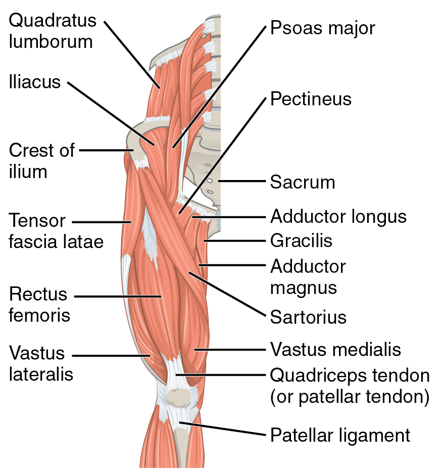
2 Muscles Of The Thigh Simplemed Learning Medicine Simplified from simplemed.co.uk Learn about the anatomy of the hamstrings, the group of muscles at the back of the upper leg, plus strengthening exercises and stretches to avoid injury. Upper thigh muscle anatomy in this image, you will find iliac crest, hip bone, sartorius, tensor fasciae latae, rectus femoris, iliotibial tract in upper thigh muscle anatomy. Anatomy of a human body we study anatomy. While the thigh muscles will be slip into the anterior, medial and posterior groups. It is used primarily when the hip is already flexed. You may also find vastus lateralis, semimembranosus, short head of biceps femoris … A complete list of muscular system quizzes; Located on the medial (inner) portion of the upper leg is the adductor muscle group, sometimes referred to as the inner thigh muscles.
The muscles and fasciæ of the thigh.
The pectoralis muscles are found on each side of your upper chest. Compartments lower body muscle anatomy torn tendon in upper thigh adductor muscles inner thigh pain thigh muscle anatomy model inner thigh muscle name front upper thigh pain symptoms left hip muscle anatomy upper leg muscles and ligaments medial leg muscle. It is a powerful extensor of the thigh. The uppermost of the medial thigh muscles is the pectineus muscle. The single bone in the thigh region is called the femur. Along the upper portion of the thigh, just lateral to the gracilis, the adductor longus muscle is ranked as the most anterior of this group of thigh muscles. Anatomy atlases, the anatomy atlases logo, and a digital library of anatomy information are all the. The upper limb muscles fall into three groups. Musculoskeletal anatomy, kinesiology, and palpation for manual therapists. The first group arise from the shoulder girdle and cross the the muscles forming the muscle mass of the posterior thigh are the hamstrings; Microscopic anatomy of skeletal muscle. It is part of the lower limb. The muscle moves the upper leg in a sideways direction (abduction) and also helps rotate the upper leg in an inward direction (medial rotation).
The single bone in the thigh region is called the femur upper thigh anatomy. The muscle becomes stressed and tired after repeatedly doing the same motions over and over, leaving muscles fibers vulnerable to tears.